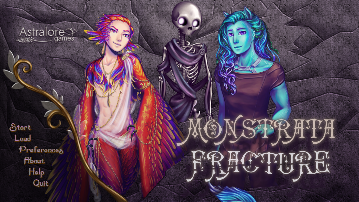
Monstrata Fracture Mac OS
Mother's Day Celebration
Save up to 35% on glass prints.
Treat Mom to a gift she’ll cherish forever with glass prints of her favorite memories. See below for details. Offer ends May 9th.
Save 25% on $75+
Fracture Walkthrough Please note that the details below reflect the time and playthroughs required to get all the Achievements in this walkthrough. Don't date monsters. Your mother would be disappointed.
- Fascia iliaca compartment block (FICB) is a volume based block used to treat pain associated with hip fractures. An anesthesia group at a central Illinois hospital expressed interest in the creation of an evidenced-based protocol for FICB. After an extensive literature search an evidence-based protocol was designed. The initial implementation of the protocol was a presentation of an.
- . Mac OS X 10.8+ (PPC not supported). Windows 8+. CPU with SSE2 support. Minimum recommended CPU: Core 2 Duo, 2GHz SETUP: 1. Unpack the FRACTURE.zip file 2. Via the FRACTUREINSTALLERS folder, run the installer for your system. A) Windows Users: please take care to.

Save 30% on $225+
Save 35% on $350+
Is there anything moms can't do?
From giving the best hugs to juggling work with kids learning from home, this year moms have taken on more than ever. Show her that you see her for the hero that she is with a thoughtful, personalized gift she’ll love.
Small Prints
Medium Prints
Large Prints
HOW IT WORKS
Fracture prints your photos directly on glass.
We’ve created a way to turn digital images into frameless glass artwork. Discover the anatomy of a Fracture print.
OUR BELIEF IN SUSTAINABILITY
We tread lightly on the planet.
Fracture is a carbon-neutral company that is always on the lookout for innovative ways to protect our planet from the impact of waste and disposable products. From glass to production to packaging, we’re committed to leaving a small footprint.
Ready to begin?
It’s easy. Find a photo you love, upload it, and pick a size. We’ll take it from there.
Share your moments.
Your photos mean the world to you. Now share them with the rest of the world.
Share your story with #FOCUSONMOMENTS.
We want you to love your prints.
We want you to be overjoyed by the photos you print. If anything in your print doesn’t meet your expectations, we’ll make it right.
What You Need to Know- Distal radius fractures are one of the most common types of bone fractures. They occur at the end of the radius bone near the wrist.
- Depending on the angle of the break, distal radius fractures can be classified into two types: Colles or Smith.
- Falls are the main cause of distal radius fractures. They may also occur during trauma from a vehicle accident or sports injury.
- Treatment varies but may include a sling or cast and sometimes surgery in the case of an unstable or displaced fracture.
What is a distal radius fracture?
The radius is one of two forearm bones and is located on the thumb side. The part of the radius connected to the wrist joint is called the distal radius. When the radius breaks near the wrist, it is called a distal radius fracture.
The break usually happens due to falling on an outstretched or flexed hand. It can also happen in a car accident, a bike accident, a skiing accident or another sports activity.
A distal radius fracture can be isolated, which means no other fractures are involved. It can also occur along with a fracture of the distal ulna (the forearm bone on the small finger side). In these cases, the injury is called a distal radius and ulna fracture.
Depending on the angle of the distal radius as it breaks, the fracture is called a Colles or Smith fracture.
- A Colles fracture may result from direct impact to the palm, like if you use your hands to break up a fall and land on the palms. The side view of a wrist after a Colles fracture is sometimes compared to the shape of a fork facing down. There is a distinct “bump” in the wrist similar to the neck of the fork. It happens because the broken end of the distal radius shifts up toward the back of the hand.
- A Smith fracture is the less common of the two. It may result from an impact to the back of the wrist, such as falling on a bent wrist. The end of the distal radius typically shifts down toward the palm side in this type of fracture. This usually makes for a distinct drop in the wrist where the longer part of the radius ends.
What are the symptoms of a distal radius fracture?
- Immediate pain with tenderness when touched
- Bruising and swelling around the wrist
- Deformity — the wrist being in an odd position
What is the treatment for a distal radius fracture?
Decisions on how to treat a distal radius fracture may depend on many factors, including:
- Fracture displacement (whether the broken bones shifted)
- Comminution (whether there are fractures in multiple places)
- Joint involvement
- Associated ulna fracture and injury to the median nerve
- Whether it is the dominant hand
- Your occupation and activity level
In any case, the immediate fracture treatment is the application of a splint for comfort and pain control. If the fracture is displaced, it is reduced (put back into the correct position) before it is placed in a splint. Fracture reduction is performed under local anesthesia, which means only the painful area is numbed.
Nonsurgical Treatment
If the distal radius fracture is in a good position, a splint or cast is applied. It often serves as a final treatment until the bone heals. Usually a cast will remain on for up to six weeks. Then you will be given a removable wrist splint to wear for comfort and support. Once the cast is removed, you can start physical therapy to regain proper wrist function and strength.
X-rays may be taken at three weeks and then at six weeks if the fracture was reduced or thought to be unstable. They may be taken less often if the fracture was not reduced and thought to be stable.
A displaced fracture needs to be corrected first. Once it is anatomically aligned, a plaster splint or cast is applied. The reduction (closed reduction) is usually performed with local anesthesia. Your orthopaedic surgeon will evaluate the fracture and decide whether you will need surgery or if the fracture can be treated with a cast for six weeks.
Surgery for Distal Radius Fractures
This option is usually for fractures that are considered unstable or can’t be treated with a cast. Surgery is typically performed through an incision over the volar aspect of your wrist (where you feel your pulse). This allows full access to the break. The pieces are put together and held in place with one or more plates and screws.
Monstrata Fracture Mac Os Download
In certain cases, a second incision is required on the back side of your wrist to re-establish the anatomy. Plates and screws will be used to hold the pieces in place. If there are multiple bone pieces, fixation with plates and screws may not be possible. In these cases, an external fixator with or without additional wires may be used to secure the fracture. With an external fixator, most of the hardware remains outside of the body.
Monstrata Fracture Mac Os 11
After the surgery, a splint will be placed for two weeks until your first follow-up visit. At that time, the splint will be removed and exchanged with a removable wrist splint. You will have to wear it for four weeks. You will start your physical therapy to regain wrist function and strength after your first clinic visit. Six weeks after your surgery, you may stop wearing the removable splint. You should continue the exercises prescribed by your surgeon and therapist. Early motion is key to achieving the best recovery after surgery.